New Research Study Demonstrates Applicability of 3-D Cell-CT™ Imaging and Analysis Platform
3D Cellular Imaging Represents an Important New Diagnostic Technology
SEATTLE, WA (December 11, 2017) – New research conducted by Arizona State University’s (ASU) Biodesign Institute and published in Science Advances (published by the American Association for the Advancement of Science, AAAS) revealed for the first time the application of 3-Dimensional Optical Computed Tomography on the cellular imaging of live cells. Read more about the ASU study and accompanying press release.
This important study further demonstrates that the 3D Optical CT approach utilized by VisionGate’s CellCT™ Imaging and Analysis Platform provides a significant advantage over other 3D imaging modalities that are based on conventional 2D microscopy, and the emergence of live cell CT adds the fourth dimension of time. The study’s principal investigator, Prof. Deirdre Meldrum, predicts that “the Cell-CT platform combined with live cell imaging and fluorescence marker analysis will revolutionize our understanding of disease at the cellular level where disease prediction and companion diagnostics for personalized medicine are poised to change the paradigm for improved health care.”
Moreover, this validates that the principles of the Cell-CT technology can be expanded into the realm of live cell imaging and analysis. This technology was originally developed by VisionGate, and the company is nearing commercialization of the Cell-CT platform for the detection of early stage lung cancer.
VisionGate’s evolving capability to perform true 3-dimensional imaging at the single cell level holds the promise to enable better understanding of how cells function, opening the door for development of new diagnostic methods and ultimately the development of new medicines.
For more information on VisionGate, please visit www.visiongate3d.com.
About VisionGate®
VisionGate is a clinical stage oncology pharmaceutical and diagnostics company focused on the early detection and prevention of cancer. Our lead investigative pharmaceutical drug is oral iloprost, currently in clinical development for the treatment of pre-cancerous bronchial dysplasia and the prevention of lung cancer following a successful Phase 2 clinical trial.
The LuCED® lung test will be the companion diagnostic for oral iloprost. VisionGate’s proprietary LuCED lung test is a non-invasive liquid biopsy diagnostic test in development for detection of early-stage lung cancer, demonstrating exquisite sensitivity and specificity in blinded clinical studies. This non-invasive sputum test is processed on the world’s first automated 3D single cell imaging and analysis technology, the Cell-CT platform, named aptly because it is similar in principle to taking a CT scan of individual cells, but using visible light without harmful radiation.
With 168 issued patents in 13 countries, VisionGate expects to play a leading role in the battle against lung cancer – the world’s number one cancer killer. VisionGate, Inc. is led by Dr. Alan Nelson, physicist, bioengineer and serial entrepreneur who previously developed the world’s first and only automated screening test to detect cervical cancer, marketed globally today as FocalPoint by Becton Dickinson.
About the Cell-CT™ 3D Imaging Platform
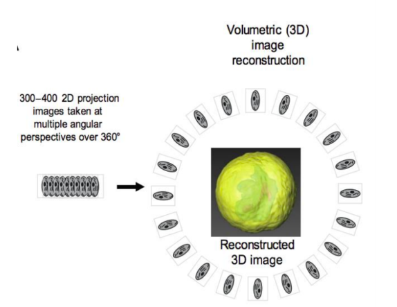
The automated Cell-CT™ 3-Dimensional Single Cell Imaging and Analysis Platform is the enabling technology which produces high-resolution 3D images of individual cells using a technique called optical computed tomography.
This 3D optical CT platform breaks new ground in the field of quantitative cell analysis by its unique ability to compute the true 3D internal structure of cells based on molecular optical absorption densities. The Cell-CT platform produces high-resolution 3D images of individual cells and measures hundreds of critical disease indicators in each cell. Together, these produce a cell classification that aids in the prediction of disease. Cells are not placed on slides, but rather, they are suspended in fluid and injected through a micro-capillary tube that permits multiple viewing perspectives around 360°.
The Cell-CT platform is a device under development and not currently cleared in the US.
Adenocarcinoma Cell as imaged by the Cell-CT™ Platform
At left – maximum intensity projection; center and right – color and opacity rendered

Adenocarcinoma Imaged by the 3-Dimensional Optical Cell-CT Platform, a Single Cell Imaging and Analysis Platform by VisionGate, Inc.
At left: maximum intensity projection single cell image; At center and right: color and opacity rendered single cell images.

Illustration of Volumetric 3D Image Reconstruction by the Cell-CT Platform.
The CellCT platform take 500 2-dimensional pictures of a 360 rotation of a single cell and then reconstruct those images into a highly detailed 3-dimensional image for analysis.


