VisionGate Presents at the World Conference on Lung Cancer for 2022 in Vienna: New Assay for Smoking Status Based on 3D Imaging of Pulmonary Macrophages

WCLC 2022 – Smoking Index
New Assay for Smoking Status based on 3D Imaging of Pulmonary Macrophages
Control # 2022-RA-152-WCLC
Meyer, Hayenga, Wilbur, Nelson
Introduction
The Cell-CTTM platform images cells in 3D with isometric, sub-micron resolution and
measures orientation invariant 3D features in each cell. The Cell-CT is being used to
identify abnormal pulmonary epithelial cells in sputum to indicate early-stage lung
cancer*. Sputum also contains pulmonary macrophages that ingest inhaled
contaminants from smoking. We hypothesize that the Cell-CT with machine learning
would discover subtle phenotypic characteristics in macrophages to differentiate current
and never smokers. Using sputum from patients without lung cancer and cells identified
by the macrophage detection classifier, we developed a second classifier to produce the
smoking index to discriminate macrophages from current and non-smoking patients.
Methods
Using a supervised learning approach, we trained the macrophage detection classifier
with 503 cell features based on 3D imaged cells and cytologically identified macrophages
(N=5,320) and non-macrophage cells (N=12,067). Focusing on sputum from 58 smoking
and 10 non-smoking patients without lung cancer, we identified 10,116 macrophages.
Cell-CT feature data from these cells was used to train a second classifier using smoking
status as ground truth to produce the smoking index probability for each cell. The smoking
index is the median of the smoking index probability for all macrophages in each case.
The smoking index was tested by identifying 202,271 macrophages from 61 current, 41
former, and 16 never smokers without lung cancer.
Results
• The macrophage detection classifier identifies cytology confirmed macrophages with
99% specificity.
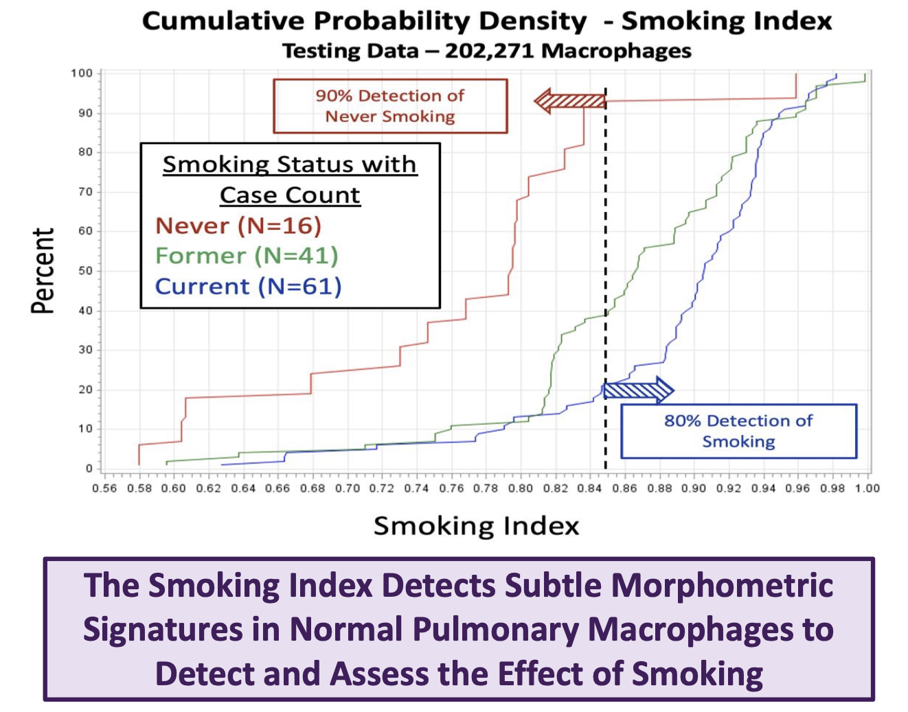
• The smoking index identifies smokers and never smokers with 80% and 90%
sensitivity, respectively. The figure shows the cumulative probability functions of the
smoking index for testing data for never (red), former (green), and current (blue)
smokers. Consider a threshold of 0.85: 80% of the smoking population has an index
above, while 90% of the never smokers have an index below this threshold,
demonstrating the robust accuracy of the smoking index.
Conclusions
This analysis demonstrates the efficacy of 3D cell analysis to characterize subtle
changes in macrophage morphology to assess smoking status. The study also suggests
a partial regression of macrophage status toward the never-smoking state following
smoking cessation. This assay is fully automated and does not require human cytology
interpretation. With further clinical studies, it may be possible to gauge the effect of
smoking and monitor smoking cessation based on the smoking index.
*Wilbur D, et. al. 2015. Automated 3-dimensional morphologic analysis of sputum specimens for lung cancer detection:
performance characteristics support use in lung cancer screening. Cancer Cytopathology 123: 548-556.


